When healthcare professionals need to examine structures inside the human body, they often rely on diagnostic imaging tests. These imaging techniques allow physicians to identify injuries, diseases, and abnormalities that may not be visible during a physical examination. Among the most commonly used imaging methods are X-rays, Computed Tomography (CT) scans, and Magnetic Resonance Imaging (MRI). As Kasey McKillip mentions, each of these technologies uses a different approach to create images and serves specific diagnostic purposes.
Kasey McKillip Briefly Highlights the Differences Between X-Ray, CT scan, and MRI
X-rays are among the most frequently performed diagnostic imaging procedures and are often the first test ordered when a patient presents with certain symptoms or injuries. This imaging method uses a controlled amount of radiation to produce images of the body’s internal structures.
A CT scan represents a more advanced form of X-ray imaging. This technology combines multiple X-ray images taken from various angles around the body to create highly detailed cross-sectional views. Using sophisticated computer processing, these images are assembled into a comprehensive three-dimensional representation of the area being examined. In some cases, a contrast agent may be administered to improve visualization of blood vessels, organs, and other internal structures
One of the primary differences between X-rays and CT scans is the level of detail they provide. While both technologies use radiation, X-rays generate two-dimensional images, whereas CT scans create three-dimensional views by combining numerous cross-sectional images. As a result, CT scans can reveal injuries and abnormalities involving internal organs and soft tissues that may not be visible on standard X-rays. Although X-rays remain highly effective for identifying fractures and certain diseases, CT scans offer a more comprehensive assessment of complex medical conditions.
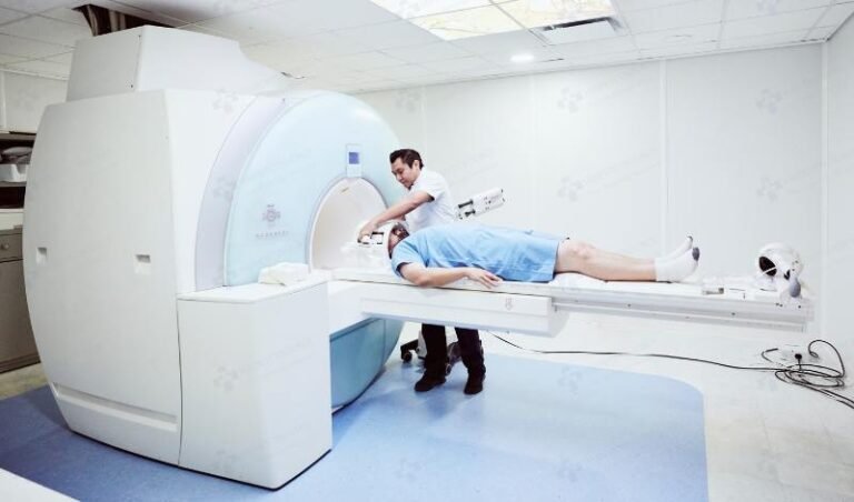
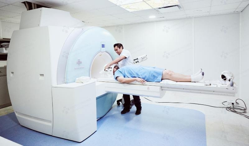
MRI, or Magnetic Resonance Imaging, utilizes an entirely different technology. Instead of radiation, MRI employs powerful magnetic fields and radio waves to generate detailed images of the body’s internal structures. During the examination, magnetic fields align particles within the body, while radiofrequency signals create responses that are processed by a computer into highly detailed cross-sectional images.
The distinction between CT scans and MRI examinations is pretty significant. In the opinion of Kasey McKillip, while procedures generate highly detailed images, CT scans use ionizing radiation, while MRI relies on magnetic fields and radio waves. Another notable difference is examination time. CT scans are generally completed within a few minutes, making them particularly useful in emergency situations. MRI examinations typically require a longer scanning period, often lasting 30 minutes or more, depending on the body area being evaluated and the complexity of the study.
Comparing MRI directly with X-rays highlights additional differences. While X-rays can be completed within minutes and are highly effective for evaluating bone injuries, MRI offers much greater versatility. MRI examinations provide detailed images of soft tissues and can identify abnormalities that may not be visible on conventional X-rays. Conditions such as ligament tears, tendon injuries, spinal cord abnormalities, and certain tumors are often more accurately assessed using MRI technology.
By selecting the most appropriate imaging modality for a particular medical condition, healthcare professionals can obtain accurate diagnostic information and develop effective treatment plans that support optimal patient care.
About the Author